
In the first three articles of this series, I’ve introduced some of my old acquaintances on Norfolk Island’s reef, celebrated those that have grown or recovered, and looked at the extraordinary damage caused by storms and powerful surges. This time, we’re looking at something rather less encouraging – coral disease and what happens afterwards.
One of the things I began noticing relatively early in my underwater photographic journey was the appearance of white patches on otherwise living corals. Sometimes they were small, but on other occasions they spread alarmingly quickly.
These weren’t necessarily bleached corals. With bleaching, the coral may remain alive despite losing much of its colour. But with white syndrome, the living tissue actually dies, exposing the white skeleton beneath it. That skeleton doesn’t stay white for long as algae and other organisms quickly move in.
Nor are all coral diseases the same. I’ve also documented a condition affecting Montipora where the coral develops a greyish-black appearance, sometimes looking almost as though it is dissolving. I wrote about this in Combine bacteria, fungi, and maybe a sponge = one toxic mess, after discussing the photographs with coral health researcher Associate Professor Tracy Ainsworth.
And this isn’t simply something I’ve noticed with my camera. Research by Page and colleagues, published in 2023, documented disease affecting around 60% of the Montipora community surveyed in Norfolk Island’s lagoon during 2020–2021. The researchers also found an association between disease occurrence and exposure to poor water quality.
I’ve photographed quite a few examples of tissue loss over the years. Some colonies have survived with scars, others have lost substantial portions of their living tissue, and some have died altogether. Not every case has an established diagnosis.
But what I’ve found particularly interesting, and concerning, is what happens afterwards. The exposed white skeleton doesn’t stay white for long. Algae and other organisms move in, and within a few years, what was once a magnificent living coral may be little more than an algae-covered skeleton. Someone swimming past today might barely give it a second glance, with no idea what was once there. Fortunately, I have the earlier photographs.
A flowerpot coral succumbs to disease
7 May 2023
22 October 2026
This is a flowerpot coral I’ve followed for several years, although its problems were already apparent when I photographed it in May 2023.
Areas of white skeleton were visible where living tissue had been lost, while much of the remaining colony was still alive, with its characteristic extended polyps surrounding the affected areas. When I showed the photographs to coral health researcher Associate Professor Tracy Ainsworth, she suggested it could possibly be black-band disease.
By October 2026, much of the once-living surface was colonised by other organisms.
I photographed this colony on numerous occasions between those dates, and included some of the intermediate photographs in an earlier post, Warning signs: quiet and unnoticed collapse of two coral colonies.
And it’s a rather sad history.
Six years of ups and downs
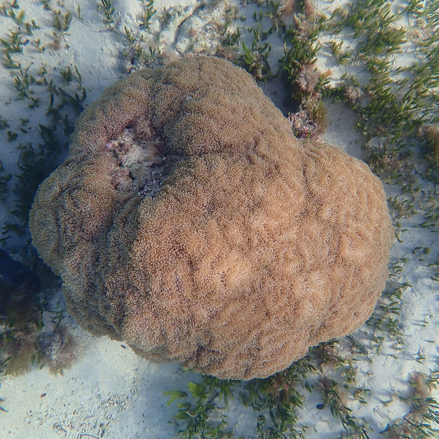
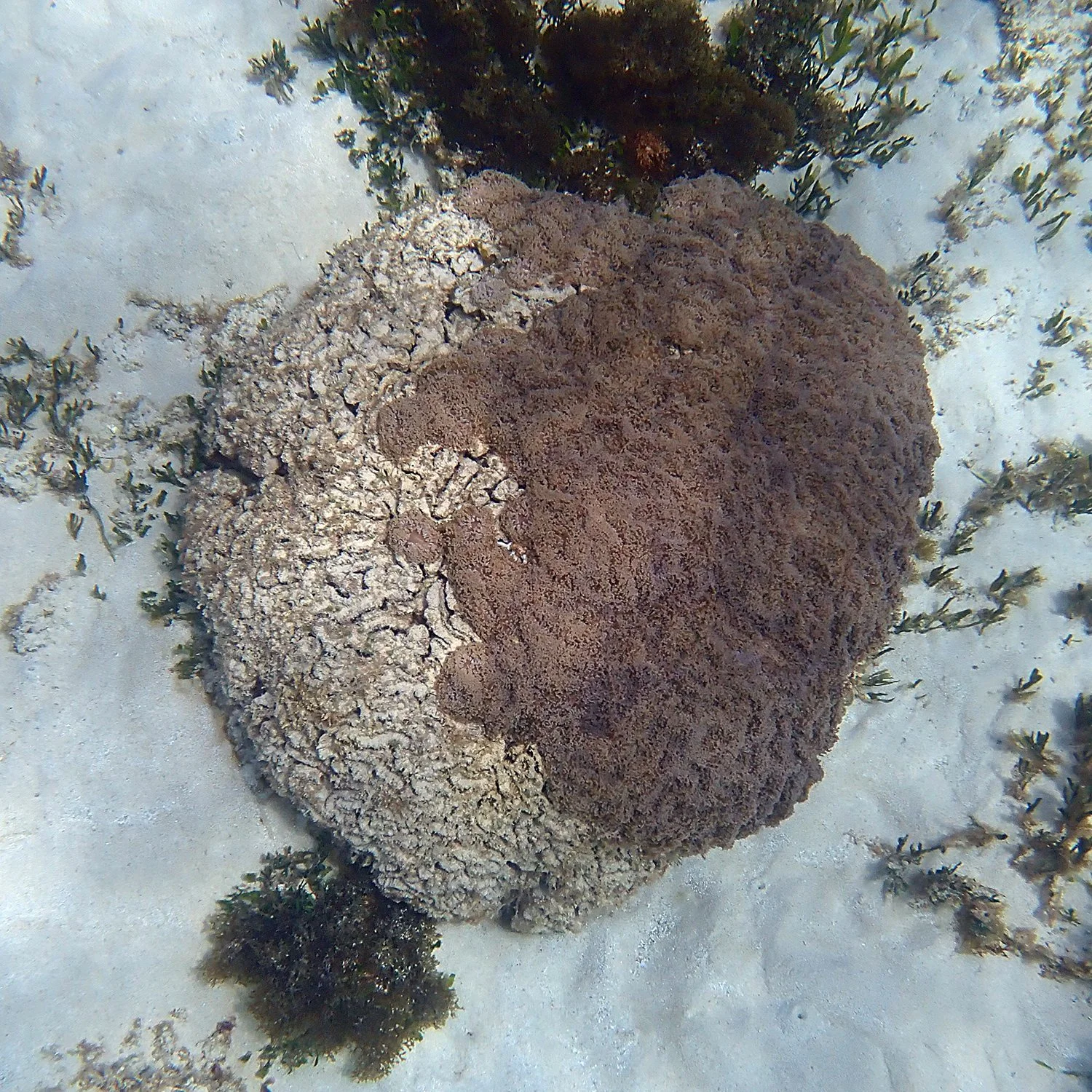
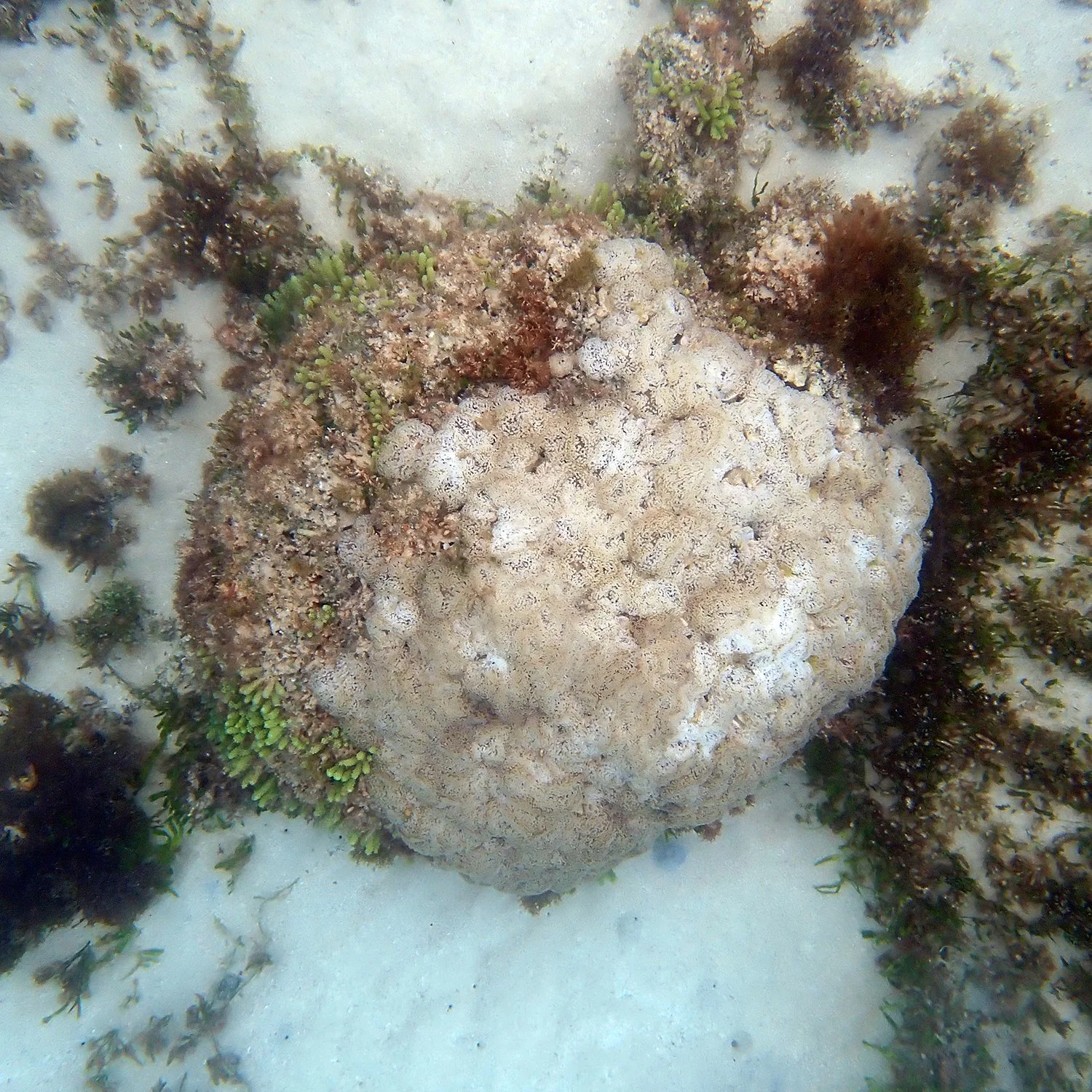
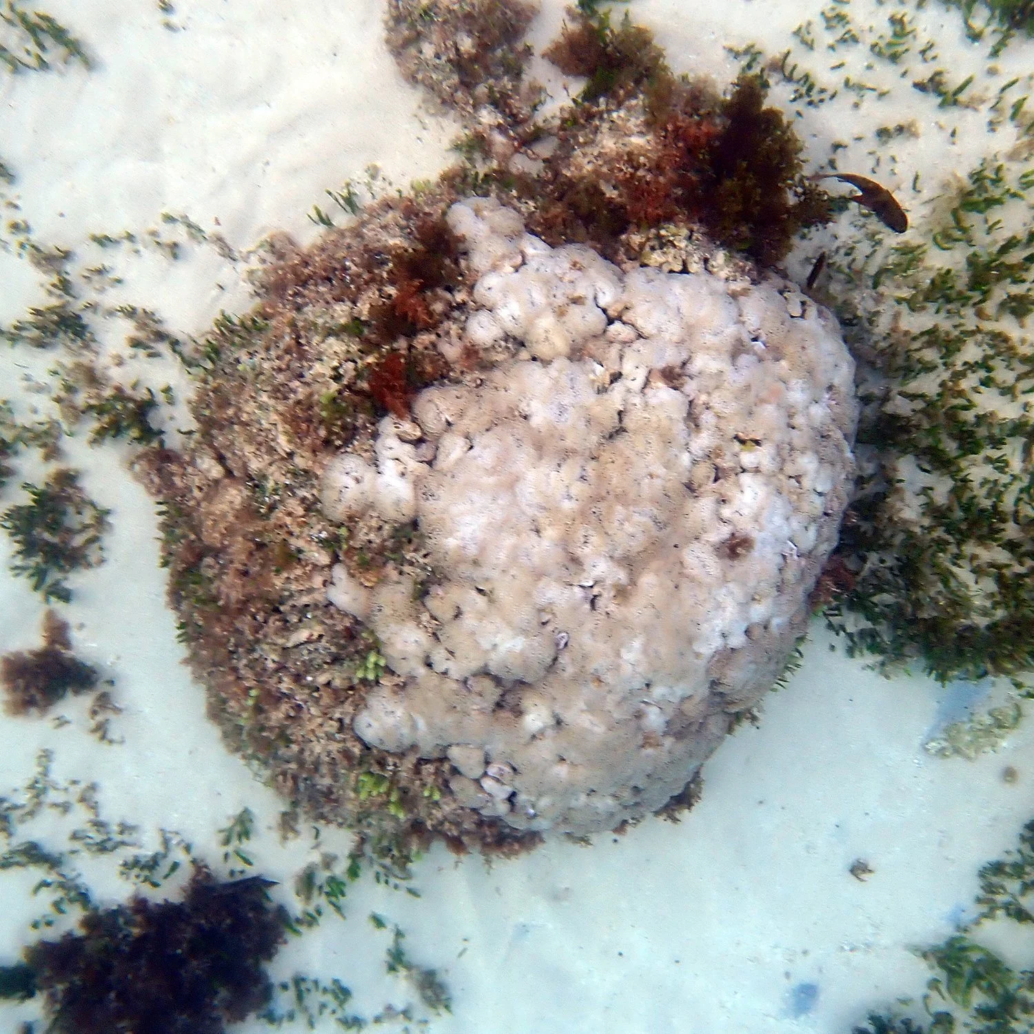
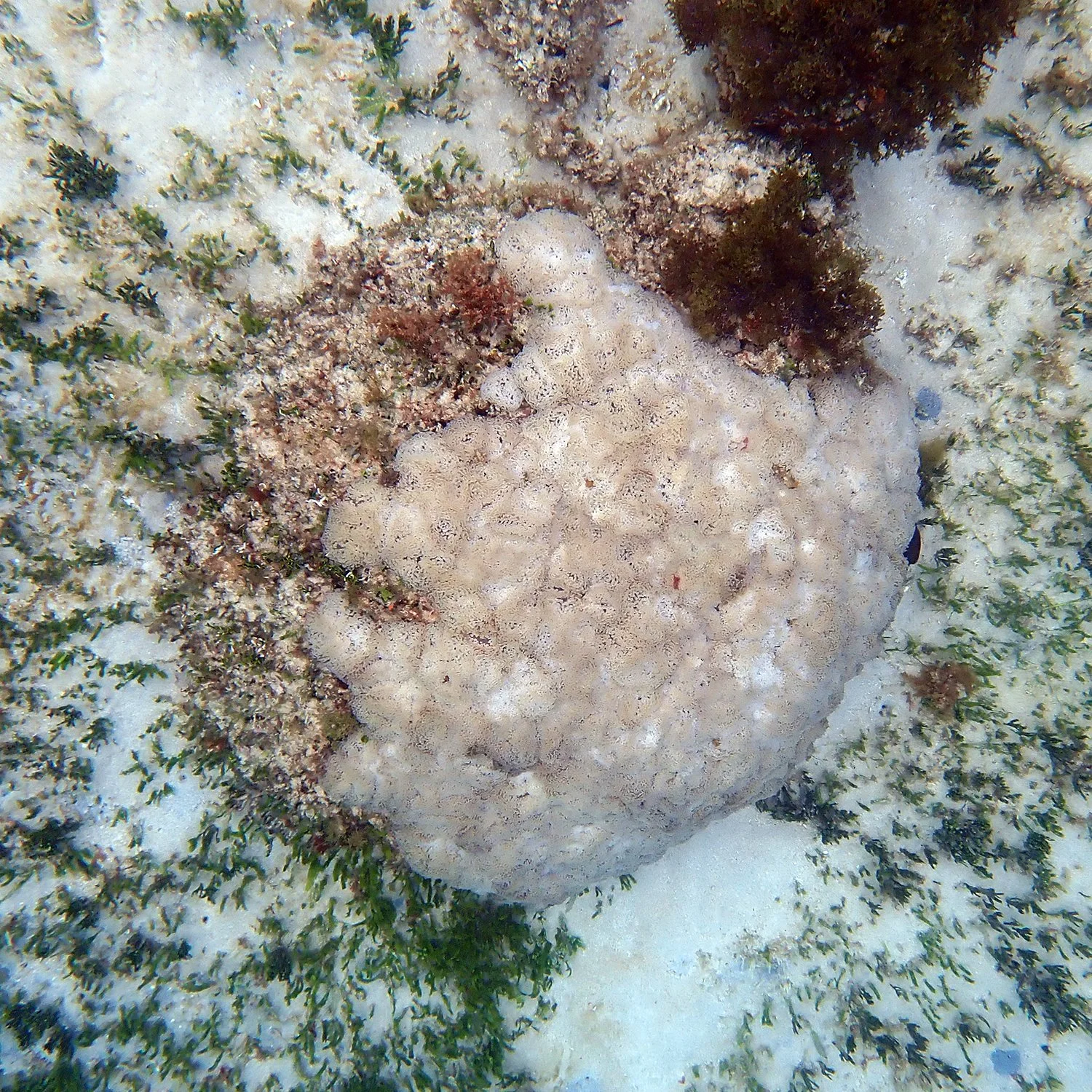
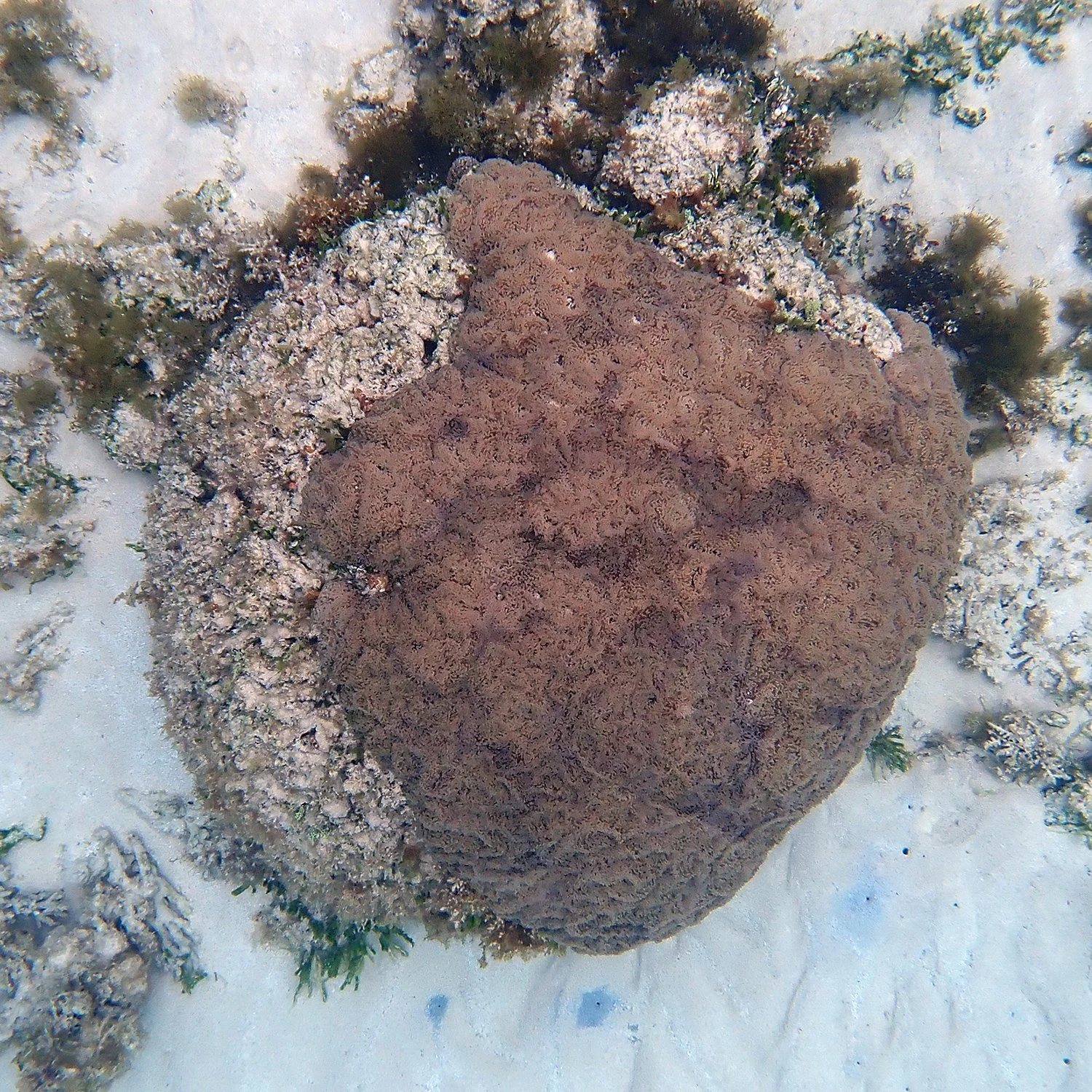
Here’s another flowerpot coral that has been part of my photographic collection almost since the beginning. I originally photographed it in July 2020 simply because I liked it. There was nothing particularly scientific about my choice!
Over the years, I’ve watched it wax and wane. There have been periods of bleaching and subsequent recovery, when the coral regained its colour and appeared to be doing rather better. I’ve included some photographs taken along the way, showing just how much its appearance has changed.
But despite surviving earlier episodes of stress, by September 2026 the colony had lost a substantial amount of its living tissue, although it had gained some from the lows of August 2022.
What interests me about this one is that its history hasn’t been a steady progression from healthy to sick to dead. It has experienced setbacks and recoveries along the way, sometimes looking decidedly worse for wear before apparently bouncing back.
Another familiar face disappears
26 January 2024, Paragoniastrea australensis
28 August 2026
This Paragoniastrea was already showing conspicuous areas of tissue loss when I photographed it in January 2024. Coral health researchers thought it might be blackline disease.
The colony was approximately 60 cm across (I know, because I used my husband’s building tape measure to check it out and wrecked it!). These brain corals are remarkably slow-growing, and this one could have been many decades old, possibly even a century. Yet in fewer than 100 days, between January and early April 2024, most of its living tissue had disappeared.
I photographed the progression repeatedly during those three months, and you can see the extraordinary sequence in my earlier article, One hundred year-old coral gone in less than one hundred days.
When I returned in August 2026 to see how it was faring, only a small remnant of living coral tissue remained at the very top. Instead, the familiar rounded structure remained, with algae and other organisms growing across most of its surface.
So, a coral that may have taken the better part of a century to grow lost most of its living tissue in little more than three months. And someone swimming past today might not even recognise it as the remains of a once-living coral colony.
When the Montipora disappears
26 February 2023
9 August 2026
This sequence shows an extensive assemblage of Montipora affected by white syndrome.
In February 2023, conspicuous white patches were visible across the living coral. By August 2026, the scene had changed considerably. Much of the original Montipora cover had been lost, and green Caulerpa was growing through parts of the affected area.
There’s an interesting seasonal story here, too. Caulerpa can grow quite spectacularly at certain times of the year and then die back. I’ll explore that in more detail in the next article, ‘When algae move in’. For now, compare how much living Montipora occupied this area in 2023 with what remained three and a half years later.
A fast-moving disease with a long aftermath
23 November 2023
9 December 2023
22 August 2026
This Hydnophora pilosa colony in the channel between Emily and Slaughter Bays is one I’ve documented in considerable detail.
On 23 November 2023, I noticed a new area of bright white skeleton where living tissue had recently been lost. Over the following weeks, I returned repeatedly, photographing the advancing white syndrome as it spread across the colony. Within a relatively short time, approximately a quarter of the colony had been affected. I also noticed fish, particularly multispine damselfish and banded scalyfins, nibbling around the affected tissue.
I documented the outbreak at the time in The spatiotemporal dynamics of a coral disease, including photographs showing how quickly the lesion spread.
Now look at the photograph from May 2026. The area that lost its living tissue is occupied by algae and other organisms. Over time, I’ve watched the appearance of that growth change, with different algae and cyanobacteria becoming more or less conspicuous at different times of the year. The original white lesion has long since disappeared from view, but its consequences remain.
I think this is one of the most revealing sequences I’ve collected. Someone seeing that algal-covered structure today would have little idea what happened to the coral in November and December 2023.
Four and a half years later
29 January 2022
25 September 2026
This final comparison is another one I find particularly sobering.
In January 2022, there was still extensive living Montipora across this patch of reef, although tissue loss was already apparent. By September 2026, much of that coral had disappeared. There are surviving patches of living coral, but relatively little obvious new coral growth occupying the area that was lost.
And this is what concerns me most about some of these photographic sequences. Coral tissue can disappear remarkably quickly, yet years later the same space may still have very little living coral occupying it.
I’ve documented a similar story involving an Acropora colony affected by white syndrome in late 2021. Nearly four years later, the dead skeleton remained occupied largely by algae and sea squirts, with only a tiny amount of new coral growth. You can see that sequence in The fate of a coral colony when it succumbs to white syndrome – four years on.
Looking at these comparisons, I can’t help wondering how many more years it might take for living coral to occupy those spaces again. If it does at all.
After the disease
One of the things I’ve learned from returning to these colonies over the years is that the most dramatic stage of a disease episode isn’t necessarily the end of the story.
A patch of exposed white skeleton may become covered by algae within weeks or months. Several years later, the original colony may be almost unrecognisable. Someone swimming past today may have no idea that what looks like an ordinary patch of algae-covered reef was once occupied by living coral. Which brings me to the point of this series of articles. The photographs allow us to look back and see what we have lost, and gained, over time.
Of course, not all the tissue loss I’ve photographed can be attributed to disease, and not every affected coral has died. Some survive with substantial portions of their colonies intact, and I’ve documented recoveries elsewhere. But what troubles me when looking through these particular sequences is how much living coral has disappeared, and how little living coral appears to have replaced it in some of the areas I’ve followed. The disease may have run its course, but that doesn’t mean the reef has recovered.
And what happens to the space left behind brings me rather neatly to the next article – ‘When algae move in’.
Reference
Page, C. E., Leggat, W. P., Egan, S., & Ainsworth, T. D. (2023). A coral disease outbreak highlights vulnerability of remote high-latitude lagoons to global and local stressors. iScience, 26(3), 106205.